Brushing up on oral diseases
View(s):The Joint International Conference and sessions of the Sri Lanka Dental Association will be held on June 27. Here Dr. Hemantha Amarasinghe looks at the country’s dental health challenges
Oral diseases are one of the most common health problems in any country.
Dental caries, periodontal diseases and oral cancers are the main oral health problems in Sri Lanka, according to the National Oral Health Survey (NOHS) conducted by the Health Ministry in 1984, 1994 and 2003.
According to the NOHS:
- 65 % of six-year-olds have dental decay in primary dentition
- 40 % of 12-year-old children have dental decay in permanent dentition.
- Gum diseases are very common — 76 % of 12-year-olds, 74 % of 15-year-olds, 91 % of 44 to 53-year-olds and 99 % of those above 65 years have gum diseases.
- 58 % of 12-year-olds had tartar (calculus), according to the 2003 NOHS.
Although oral diseases are the most common health problem in the country, the surveys have also indicated a declining trend in these diseases.
Oral cancer is the most common cancer in Sri Lanka among males and ranks sixth among women and accounts for 12.9 % of total malignancies in the country. The incidence of cancer of the oral cavity and oro-pharynx in Sri Lanka, standardised to the world standard population in 2006, was 16 per 1,000 and 4 per 100,000 in males and females respectively.
All oral and dental health diseases can be prevented.
Prevention of dental decay and gum diseases
Dental decay and gum diseases can be prevented by proper dietary and oral hygiene practices.
- To prevent dental decay, the frequency and amount of sticky sweet foods (toffees and chocolates) and sticky starchy food (biscuits, buns and pastries) should be reduced.
- Fresh fruit, vegetables and cereals should be eaten instead of sweets and shorteats.
- Sweets should be eaten soon after the main meals and not in-between the main meals.
- Brushing methodically twice a day — morning and night — with fluoridated toothpaste to remove plaque in the gum margin will prevent dental decay and gum diseases.
- A six-monthly check by a Dental Surgeon will ensure dental health.
Prevention of oral cancer
- Avoid betel chewing as tobacco, arecanut and lime can cause oral cancer
- Avoid tobacco in any form — cigarettes, bidi, pipe, snuff
- Avoid alcohol use
- Avoid consuming commercially prepared areca products like babul, bida, panparg, gutkha, mawa
- Improve oral hygiene by brushing the teeth and visiting the Dental Surgeon regularly.
- Eat plenty of fruit and vegetables — 5 portions per day or 400gms
Early detection of oral cancer
Oral cancer is often preceded by oral pre-cancer.
In Sri Lanka, the prevalence of oral leukoplakia and oral sub mucous fibrosis (OSF) is reported as 26.2 and 4.0 per 1000 respectively. Studies in central Sri Lanka estimated the prevalence of all OPMD at 4.2 %, and among tea estate workers at 6.7 %. According to the recent study in the Sabaragamuwa province, prevalence of OPMD was 11 % in the rural and estate sector.

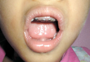
Front teeth decay in a child
The following lesions and conditions are usually described under oral pre cancer: White patch (leukoplakia), red patch (erythroplakia), submucous fibrosis, lichen planus, palatal lesions of reverse smoking, discoid lupus erythematosus, and hereditary disorders such as dyskeratosis congenita and epidermolysis bullosa. Of these, leukoplakia, submucous fibrosis and lichen planus are the commonly found disorders.
The health system has unfortunately failed to carry out systematic interventions to detect those with pre-cancer and to manage them, to prevent oral cancers. Early detection and appropriate management of pre-cancer with habit intervention is one of the main strategies needed to reduce the burden of oral cancer in Sri Lanka.
White patch (Oral Leukoplakia)
There are two main clinical types of leukoplakia: homogeneous and non-homogeneous. Homogeneous lesions are uniformly flat, thin and exhibit shallow cracks on the keratin surface. The risk of malignant transformation of this variety is relatively low.
Non-homogeneous lesions carry a much higher risk of malignant transformation and are classified as follows.
- Speckled : Mixed, white and red, but retaining predominantly white character.
- Nodular : Small polyploidy outgrowths, rounded red or white excrescences.
- Verrucous : Wrinkled or corrugated surface appearance.
- Proliferative verrucous leukoplakia presents with multiple, simultaneous leukoplakia, as the disease is visibly multimodal and frequently covers a wide area.
However, those with mixed white and red plaques should be recognised as having a higher risk status, and these are to be denoted as ‘erythroleukoplakia’.
Prevention is the key to manage oral leukoplakia. Patients must understand the risk of tobacco, arecanut and alcohol use. Management depends on the histological report and the site of the lesion. If no dysplasia is noted on a buccal mucosal biopsy, routine observation of the lesion is recommended. Patients with leukoplakial lesions of the floor of the mouth, lateral and ventral tongue, soft palate, and oropharynx should have these lesions removed completely whether microscopic examination reveals dysplasia or not.
Red patch (Erythroplakia)
Erythroplakia is defined by the World Health Organisation (WHO) as ‘any lesion of the oral mucosa that presents as bright red velvety plaques which cannot be characterised clinically or pathologically as any other definable disease’. Such lesions are less common than leukoplakia but very careful observation reveal an association of erythroplakia with early invasive oral carcinomas. Erytroplakia may also be associated with leukoplakia, as mixed red and white lesions. However, it is the red portion that is more worrisome than the white.

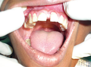
Gum disease in an adult male
Management of erythroplakia, follows the same principles of management of leukoplakia, according to its histopathological features.
Oral Submucous Fibrosis (OSF)
OSF is an important form of a potentially malignant oral condition that appears in the oral cavity. Among this special recognition was made for OSF because of its high prevalence, relatively high rate of malignant transformation than leukoplakia and possible avenues for prevention.
Worldwide estimates indicate that 2.5 million people are affected; most cases concentrated on the Indian subcontinent. A sharp spurt in the incidence of OSF was noted after packets of arecanut came onto the market and the incidence is increasing. A high prevalence of OSF (1.7 %) was reported in the Sabaragamuwa province of Sri Lanka in 2008.
OSF presents with burning sensation, blanching of the oral mucosa together with stiffness of oral mucosa and symptoms leading to restricted mouth opening, inability to protrude the tongue, altered pronunciation, and intolerance to spicy-hot foods. OSF is often misdiagnosed by general practitioners as iron deficiency anaemia, due to its pale white blanching appearance of the oral mucosa.
OSF is often resistant to treatment regimes. Submucosal injected steroids and hyaluronidase, oral iron preparations, and topical vitamin A with steroids are some of the agents that are used. All these therapies are essentially palliative. In severe cases, surgical intervention is the only treatment, but the fibrous bands and other symptoms often recur within a few months to a few years.
Oral lichen planus
Oral lichen planus is a common chronic immunologic inflammatory mucocutaneous disorder that varies in appearance on clinical presentations and is described to include both white and red components. The white forms include papular, reticular, and plaque likes lesions. The most common is the reticular form, which typically presents as a non-elevated keratotic patch with fine, lace like striations (Wickham’s striae). Red forms are less common and include atropic, ulcerative and bullous forms.
Lichen planus lesions are sometimes painful, particularly the ulcerative type. They may appear in almost any oral site but are most common on the buccal mucosa. About 28 % of patients who have oral lichen planus also have skin lesions.
Treatment is usually palliative and a small percentage of cases may predispose to the development of oral cancer.
(The writer is a Consultant in Community Dentistry attached to the National Cancer Control Programme)
Follow @timesonlinelk
comments powered by Disqus


