ECG: What it detects and what it doesn’t
 In this latest edition of our test series, Prof. Shyam Fernando, a consultant physician, explains the workings of an electrocardiogram or ECG. “An ECG is done mainly to find the cause of chest pain or palpitation,” explains Prof. Shyam. “It is also done as a routine check for insurance, visa applications and before surgery.”
Considering how common the test is, here are some of the things you should know about how it works, how it is used in diagnosis and in what circumstances an ECG may not actually give you the answers you need.
What it is:
The heart is a pump made up of muscle. Tiny electric currents that spread through the heart make the heart muscle contract in a rhythmic coordinated manner. This pumps blood through the body. The ECG records these electric currents.
How it is it done:
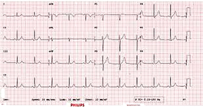
You will be asked to lie down on a couch. Small patches called electrodes will be stuck to your arms, legs and the chest. It may be necessary to shave some hair to make sure they stick to the skin. These patches are connected to a machine with wires. The machine amplifies the electrical currents that occur with each heart beat and records them on paper as a wavy line. It is called a 12-lead ECG because the machine ‘looks at the heart’ from several angles and records 12 different wave patterns.
How is it used in diagnosis?
Any damage to the heart muscle, as in case of a heart attack, can be detected on ECG. By inspecting the 12 different wave forms recorded, it is even possible to say which part of the heart is damaged. Excessively fast, slow or irregular heart rhythm, enlargement of the heart or inflammation of the heart muscle also can be detected on the ECG. Any abnormality seen on the ECG should be correlated with symptoms and findings on examination before coming to diagnostic decisions.
How do I prepare?
No special preparation is necessary. Do not exercise just before taking the ECG. It is important that you relax and lie still during the test. You may be asked to hold your breath for a few seconds. The test takes about 5 minutes. You will get the printout of the recording immediately afterwards.
What are the after-effects?
None. It is painless and harmless. You won’t get electrocuted.
What are the pitfalls?
In certain situations, the ECG might not detect even significant heart problems. In other words, a normal ECG does not always rule out heart disease. An irregular rhythm that ‘comes and goes’ might not ‘be caught’ during the five-minute period the ECG is recorded. In this sort of situation, your doctor might arrange a Holter monitoring, which is an ECG, recorded constantly throughout one whole day.
Another situation where an ECG done at rest could be perfectly normal is angina. Angina is a common heart condition, where chest pain occurs only during exertion (eg. walking) and subsides soon after stopping. This is due to ischaemia or narrowing of coronary blood vessels. Only an exercise ECG, where the recording is done while you walk on a treadmill can detect this.


