Cardiac imaging, the multi-tasker at work
View(s):A pain in the chest sends men or women rushing to the nearest hospital, for chest pain and heart attacks are synonymous.
Whereas earlier the reaction in hospitals would be to suspect ischaemic heart disease, an angiogram would be performed to check out whether the chest-pain sufferer is having coronary artery disease. This would be followed by a flurry of activity ending in bypass surgery or stenting. Now, however, there is a different and more effective gate-keeper, MediScene learns.
‘Cardiac imaging’ is now available in Sri Lanka and the pioneering Consultant Cardiologist behind this new test, a better and safer option, is 42-year-old Dr. Prakash Priyadharshan.

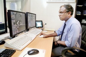
Dr. Priyadharshan performing a Cardiac CT. Pic by M.A. Pushpa Kumara
It is on a busy clinic day that MediScene meets Dr. Priyadharshan who is attached to the Institute of Cardiology of the National Hospital of Sri Lanka. Having returned after training in this specialty at Leeds and Bradford Hospitals in the United Kingdom three years ago, he had initially served at the Polonnaruwa Hospital, setting up the Cardiology Unit there.
Getting down to the nitty-gritty of this highly-skilled arena of imaging of the heart, the organ that keeps a person alive, he says cardiac imaging is “multi-tasking work” and is a current technological advance in identifying coronary artery disease.
“This is a very good test, an advanced gate-keeper between chest pain and treatment, whereas earlier there were only indiscriminate angiograms,” he says.
Dr. Priyadharshan deals with the steps that doctors would go through when a person seeks treatment for chest pain (angina). The first suspect would be a plaque build-up and a blocked blood vessel.
Pointing out that if there is plaque build-up and a blocked vessel the consequences would be poor blood supply or loss of blood supply to the heart muscle; change in muscle-cell chemicals (metabolism); and change in heart contractions, he says that the patient’s case history would indicate chest pain. Changes would also appear in an electrocardiogram (ECG).
“The person would then be instructed to undergo an Exercise ECG, which is the ‘traditional gate-keeper’. If negative, the patient would be cleared of coronary artery disease,” he says.
However, MediScene learns that not all patients can undergo an Exercise ECG, as some cannot run, others may have medical conditions which would endanger them during the test and there could also be interpretation difficulties of certain ECG changes.
The next best option has been an angiogram to check for coronary artery disease, he says, pointing out that it is not only invasive and risky but also costly and has to be performed in a Catheterization Laboratory, which in government hospitals would be scarce.
 “Doing an angiogram as a diagnostic test is a waste of time and resources when the procedure can be better utilized for treatment (therapy),” he points out.
Complications for the patient through this invasive test are not the only issue, he points out. In the state health service there would be long waiting lists for angiograms, with treatment time being wasted for diagnosis, while the maintenance of Cath Labs is also very costly.
“An angiogram would not catch borderline blocks (50-70%) which would be the cause for the symptoms of chest pain. It would also not give any information whether the heart muscle that was being supplied by the partially-blocked or blocked blood vessel was already dead and whether it was non-viable,” he says.
Such angiograms would be followed by invasive, risky and expensive procedures like coronary artery bypass grafts (CABG) or stenting, MediScene understands.
In cardiac imaging, however, it is not only the vessel that is being looked at as in an angiogram but also the muscle. “We get the whole picture,” he reiterates, explaining that the ischaemic myocardium will show lack of perfusion (blood supply), changes in metabolism, changes in contracting ability and changes in the ECG.
The accuracy of cardiac imaging is such that not only will it show where and how many blocks there are but also what the blood supply situation is and whether any area of heart muscle is already dead. Thus it will guide doctors on whether the patient needs to undergo a bypass or stenting, it is learnt.
Crucial information on the remaining heart function and the patient’s risk of suffering a massive heart attack or death will also be gleaned through cardiac imaging, he says.
In the modern world where people’s lives are fast-paced, he says there is a reluctance to undergo a battery of tests, as it is time-consuming as well as costly.
The answer lies in the “good gate-keeper” which is capable of multi-tasking while also being accurate. Bringing to mind the example of an i-Phone which can be used for communication in several ways such as talking, texting and e-mailing while also taking photographs, Dr. Priyadharshan puts cardiac imaging on a pedestal, above the exercise ECG and also the angiogram.
Dr. Priyadharshan recalls how his gurus, Dr. S. Narenthiran and Dr. Vajira Senaratne, were quite insistent that at least one of their five Senior Registrars (SRs) should take to this field. When he decided to take up this challenge, he had been at the butt-end of sympathy from his peers that he would be stuck in a specialty where his knowledge would be wasted without the necessary equipment.
“Now three more are in training in this field,” he says, pointing out with conviction that it was the “right decision”.
Cardiac imaging has bridged the vacuum of information that we don’t get from an Exercise ECG or an angiogram, says Dr. Priyadharshan, adding that in this one test there is an answer for every single question relating to angina. So, seeing is believing in cardiac imaging.
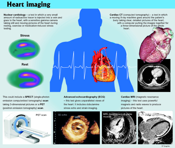
| Getting to know all that’s wrong with your heart Cardiac imaging’s superiority lies in the fact that it can be performed on a wide-range of patients, it can spot the blood-vessel block, it can see the blood supply and it can accurately predict muscle-death and also heart function, reiterates Dr. Priyadharshan. Nuclear cardiology – this test which includes SPECT (single-photon emission computerized tomography) scans and PET (positron emission tomography) scans can see the blood supply to the heart muscle. It can be performed when both the heart is resting or under stress. It is suitable for those with chest pain either in routine diagnosis or in an emergency and others who are not fit to undergo an Exercise ECG. Changes in muscle-cell chemicals come to the fore with this test. Cardiac CT (computed tomography) – this is an enhancement to visualize coronary arteries in place of a coronary angiogram. This cutting-edge imaging is used in vulnerable plaque assessment. It may be performed as an exclusion test for significant coronary artery disease, particularly in young people who have chest pain or those suffering from diabetes. Advanced Echocardiogram – this test uses high frequency sound (ultrasound) to detect disease while bounced back sound creates pictures. This is the cheapest test to pick up heart muscle contraction and other structures. Cardiac MRI (magnetic resonance imaging) – in this test, the patient is magnetized to produce an electrical signal. It can assess blood supply to the muscle and also viable muscle. |


