Sunday Times 2
Microcephaly surveillance in Lanka
View(s):Dr. Kapila Jayaratne
Microcephaly, small head size in new born, is a rare condition. This condition has recently gained much media and public focus because of its association with the Zika virus. On February 1 this year, WHO declared a Public Health Emergency of International Concern (PHEIC) in response to reports of clusters of microcephaly cases and neurological disorders in the areas affected by the Zika outbreak especially Latin America. Microcephaly is a clinical sign and not a disease.

Kapila Jayaratne
Until recently, there was no standard surveillance case definition for microcephaly. At present microcephaly is broadly defined as a small head size, typically greater than two standard deviations below normal.
To assess whether a baby has microcephaly, WHO advocates measuring head circumference 24 hours after birth, comparing the value with WHO growth standards, and continuing to measure the rate of head growth in early infancy.
The head circumference <-2SD according to INTERGROWTH standards for gestational age and sex is being preferred by many experts. The head circumference is measured via occipital frontal circumference.
Incidence: Birth defects registries in the United States have estimated that microcephaly ranges from 2 to 12 babies per 10,000 live births. Many countries, including Sri Lanka, have no valid data on microcephaly and its causality. Even estimated incidence of microcephaly has wide variation due to the differences in the definition and subjects under study.
How does it happen?
The foetus’ skull is made of six bones that are not fused at birth. The head of the baby in the womb increases in size because of the underlying brain growing. It exerts physical force on the skull bones and causes them to expand as well. The rate of expansion is the highest in the first few months and gradually plateaus over time.
There are two mechanisms for a ‘small head’ to occur; premature fusion of cranial sutures (i.e., craniosynostosis) or poor brain growth;

At present microcephaly is broadly defined as a small head size, typically greater than two standard deviations below normal.
1. If the sutures between skull bones fuse prematurely, the head circumference will grow at a much slower rate and resulting in an abnormal shape – Craniosynostosis.
2. When the foetal brain has a lower volume than normal, it may not grow as expected with normal brains. Decreased brain growth exerts decreased outward force exerted on the skull, and decreased expansion of the head circumference. This process may result in a normally shaped or symmetric head.
Microcephaly can be present at various ages and can be either static or progressive, depending on the cause.
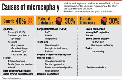
Causes of microcephaly
Various aetiologies can lead to decreased brain volume and many potential causes of microcephaly, but often the cause remains unknown.
The most common causes are given in the table:
As such microcephaly has many causes, of which Zika is potentially just one apart from viruses rubella, Cytomegalovirus (CMV) and lymphocytic choriomeningitis virus (LCMV).
The US Centers for Disease Control (CDC) announced Zika virus is the cause of a surge in birth defects across Latin America and the Caribbean, on April 13, linking Zika infection during pregnancy not only to microcephaly but also to congenital blindness, stillbirth, and other fetal abnormalities. (British Medical Journal -BMJ Published 15 April 2016). CDC says, “There is no longer any doubt that Zika causes microcephaly. We believe the microcephaly is likely to be part of a range of birth defects.” The CDC remarked, “Never before in history has there been a situation where a bite from a mosquito can result in a devastating malformation.”
Zika virus is transmitted primarily by Aedes mosquitoes. This is the same mosquito that transmits dengue, chikungunya and yellow fever. Persons with Zika virus disease can have mild fever, skin rash, conjunctivitis, muscle and joint pain, malaise or headache.
 Zika virus is primarily transmitted to people through bite of an infected mosquito. Evidence is emerging of sexual transmission of Zika virus even up to six months.
Diagnosis
During the pregnancy, early diagnosis of microcephaly can sometimes be made by foetal ultrasound. The possibility of diagnosis is higher if ultrasounds are made at the end of the second trimester, around 28 weeks, or in the third trimester of pregnancy.
Often diagnosis is made at birth or at a later stage by measurement of head circumference.
For the confirmation of cause of microcephaly various laboratory investigations and imaging tests need to be done both in the mother and the baby;
- Karyotype
- Serology/molecular testing for Zika, CMV, Rubella, toxoplasmosis, LCMV
- Cranial USS
- Histopathological study
Microcephaly surveillance
Sri Lanka is considered a country of possible risk for Zika virus transmission due to the presence of transmitting mosquito.
After WHO declared a Public Health Emergency of International Concern February 2016, and WHO advice on surveillance for microcephaly and GBS to be standardised and enhanced, particularly in areas of known Zika virus transmission and areas at risk of such transmission, an expert working group including community physicians, paediatricians, neonatologists, geneticists, virologists and administrators, emphasised the need of microcephaly surveillance in neonates and developed a surveillance format.
The Ministry of Health Sri Lanka initiated microcephaly surveillance from the second week of February this year throughout the country.
(The writer is a Consultant Community Physician. He is also the National Programme manager for maternal & child morbidity and mortality surveillance at the Family Health Bureau of the Ministry of Health. He also heads the Perinatal Society of Sri Lanka.)

