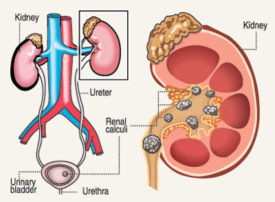
A painful saga of stones in the kidney
Renal stones (or renal calculi) form within the kidney. In subjects susceptible to develop renal stones, renal stone forming crystals pass out in the urine. And, in stone formers, renal stones, small in size, would pass out in the urine. When passing out a tiny renal stone in the urine, one may or may not feel a pain. But, a sizeable renal stone traversing the ureter or the urethra would cause intractable pain.
 A renal stone keeps on growing within the kidney, destroying the kidney matter, subsequently impairing kidney function (renal function). Medical research and experiential evidence of urologists provide proof that renal stone formation and recurrence can be halted by combined interventions: behavioural, dietary and therapeutic.
Renal stone formation (urolithiasis) is preventable. Hence, the adage “once a stone former – always a stone former” deserves a facelift.This article intends to provide guidance particularly to the following four categories of individuals:
(a) subjects prone to develop renal stone disease,
(b) patients with diagnosed renal stone disease,
(c) patients under treatment to prevent recurrence of renal stone disease,
(d) patients whose renal stones had been removed by some form of Intervention.
Intractable pain – an accompaniment of renal stone disease:
The pain referred to as ‘renal colic’ occurs when a sizeable renal stone blocks the kidney / ureter / bladder / urethra.
The pain originates below the ribcage. This pain radiates downwards along the affected side of the body from the loin area to the groin area, becoming intense and unbearable! It comes in wave-form, persisting and subsiding, repeatedly. Each pain episode can last from quarter of an hour to an hour or so.
Although ‘pain relievers’ for renal colic can relieve the pain briskly, you must know that pain suppression is only a temporary measure, not a permanent cure. The answer lies in medical intervention on the one hand and preventive commitment on the part of the sufferers on the other hand. If the preventive aspects are ignored the adage ‘Once a stone former – Always a stone former’ could never be negated. Therefore, a subject predisposed to urolithiasis and wanting to avert the gruesome episodes of renal colic and subsequent loss of kidney function (renal impairment) must think wisely.
The urine ‘Lithogenic Test’
It is apt that a subject with a family history of ‘renal stone disease’ (urolithiasis-prone) or with a likelihood of developing renal stone disease (predisposed to) arrives at a definitive conclusion – is my urine lithogenic or non-lithogenic? The Urine Lithogenic Test enables one to arrive at a definitive conclusion. If the urine is lithogenic, then the next aspect that needs unravelling is what is /are the lithogenic constituent/s in the urine? Here too, the Urine Lithogenic Test is capable of unravelling the lithogenic constituent/s in the urine. Further, once on treatment, the Urine Lithogenic Test is capable of ascertaining the success / failure, compliance / non-compliance, with regards to the treatment.
The Urine Lithogenic Test is a new medical laboratory test done by Medical Laboratory Technicians with a training to perform the test competently. The test requires a ‘mid-stream’ sample of urine collected from the ‘first morning void’. The sample of urine is collected ideally in a (clean – sterile) ‘uricontainer’ purchased from a pharmacy. A patient collecting the ‘first morning void’ must collect the urine urinated following waking up, ideally before eating food or drinking fluids. A ‘mid-stream’ sample of the urine is obtained by passing out some urine (into the toilet bowl / urinal) uncollected and then collecting the urine next expelled. The uricontainer can be almost completely filled with the urine.
The urine sample thus collected must be handed over to the medical testing laboratory within 2-hours, with a request for the ‘Urine Lithogenic Test’. The laboratory requires over 24 hours to complete the Urine Lithogenic Test, review the findings and compile their report.
The Laboratory Report will contain the following information:
(a) Urine Specific Gravity (with the Laboratory Established Reference Interval Range),
(b)Whether the urine is non-lithogenic or lithogenic,
(c) If the urine is likely to be lithogenic or definitively lithogenic, the chemical identity of the lithogenic constituent/ constituents.
The information disclosed in the Urine Lithogenic Test Report will serve as a (a) guide to behavioural, nutritional and medical steps to be instituted. Further, the Urine Lithogenic Test Report shall serve to determine (b) patient compliance and (c) efficacy of treatment to avert recurrence of renal colic and also inhibition of growth of renal calculi.
(The writer is a Retired Lecturer in Clinical Biochemistry & Nutrition – Peradeniya University -Medical Faculty)
| Reasons for Calcium Oxalate renal stone formation: The principal chemical constituent of the commonest type of renal stone is Calcium Oxalate. In fact, Calcium Oxalate renal stone formers exceed 75% – 80% of the world’s renal stone disease population. The factors underlying the genesis of Calcium Oxalate renal stone are basically (a) aberrant behavioural pattern and (b) dietary aspects, both of which are adjustable or correctable.In Sri Lanka, renal stone disease is particularly prevalent in areas affiliated to Kurunegala District, Anuradhapura District, Polonnaruwa District, more than in the other areas of the island. Why? In these areas, workers engaged in intensive physical labour, are exposed to dry heat of the sun, do not drink adequate amounts of water to compensate body water loss through heavy perspiration and unknowingly form concentrated urine. Thus, the urine Specific Gravity elevates (i.e. way above 1.010). Excessively concentrated urine could contain higher levels of insoluble Calcium Oxalate, which would give rise to Calcium Oxalate crystals. In the long-term, these Calcium Oxalate crystals would coalesce to form Calcium Oxalate stones within the kidneys.Another contributory factor in the above districts was found to be due to high levels of fluoride in well water. I recall a family (i.e. mother, father, daughter) in Anuradhapura District with renal calculi seeded in kidneys, referred to me by the then Consultant Urologist of the GH/Kandy. This family had used well-water for cooking and drinking. When this water was tested it became evident that the fluoride levels were high (F‑>4ppm). Research studies in other countries also fortify the association between renal stone disease and high fluoride content in water consumed. Therefore, it makes sense to reduce the fluoride content of water for consumption to safer levels to avert renal stone disease. | |


