Trouble in the largest joint in the body
 What do a clergyman, a housemaid and a jumper have in common?
All these people and more can come down with ‘knee issues’ if they are not careful.
This is what Senior Consultant Orthopaedic Surgeon, Dr. Narendra Pinto tells MediScene, lifting up the human knee for a close look, pointing out its parts and then explaining how these parts can be affected in different ways.
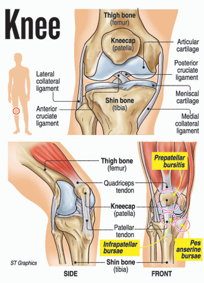
Explaining that the knee is the largest joint in the body, he says that its anatomy comprises bones, cartilage, ligaments and tendons.
Bones – the knee joint is formed when the three bones – the thigh bone (femur), the shin bone (tibia) and the kneecap (patella) meet.
Cartilage – the ends of the femur and tibia and the back of the patella are covered by the articular cartilage, which is a slippery substance which helps the knee bones to glide smoothly across each other, allowing the bending or straightening of the leg. The articular cartilage’s surface is composed of a hyaline cartilage, which is specific to the knee-joint.
Meanwhile, two wedge-shaped pieces of the meniscus cartilage act as ‘shock absorbers’ between the femur and the tibia. Unlike the articular cartilage, the meniscus is tough and rubbery and cushions and stabilizes the joint.
Ligaments – these are like four strong ropes which connect bones to other bones to keep the knee stable. The ‘collateral’ ligaments on the sides of the knee are the ‘medial collateral’ ligament on the inner side of the knee and the ‘lateral collateral’ ligament on the outer side of the knee control the sideways motion of the knee and brace it against unusual movement.
The ‘cruciate’ ligaments inside the knee-joint which cross each other to form an ‘X’ with the ‘anterior cruciate’ ligament in front and the ‘posterior cruciate’ ligament at the back control the back and forth motion of the knee.
Tendons – the muscles are connected to bones by tendons. The quadriceps tendon connects the muscles in the front of the thigh to the patella, while stretching from the patella to the shin bone is the patellar tendon.
Dr. Pinto points out that the knee also has up to 11 bursae or flattened, thin fluid-filled sacs which are under the skin over the knee-joint. They are like cushions between the tendons and the bones. These bursae, filled with synovial fluid (the body’s natural lubricating fluid), help different tissues such as muscles, tendons and skin to glide and slide over the bony points such as the kneecap, reducing friction among them.
He picks out bursitis as these bursae can become irritated, inflamed and swollen and points out that this condition commonly affects the prepatellar bursae (which lie in front of the patella); the infrapatellar bursae (which lie underneath the patella); and the Pes anserine bursae 9on the inner side of the knee). The swelling is superficial but it is a very painful condition.
The causes of bursitis, according to Dr. Pinto, include overuse; a direct blow (trauma) to the knee may be when engaging in a sport, chronic friction or repetitive action such as frequent kneeling or over-exercising and ageing.
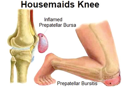
Housemaid’s knee – this is when the prepatellar bursae located just in front of the kneecap beneath the skin get inflamed. Prepatellar bursitis is dubbed housemaid’s knee because maids in those days were on their knees to clean the floor. It can affect anyone who is on his/her knees for long periods of time and causes pain and swelling at the front of the knee.
 While volleyball players can get this condition by diving onto their knees for the ball, wrestlers may also suffer the same fate when their knees rub against the mat constantly.
Clergyman’s knee – this is infrapatellar bursitis when the infrapatellar bursae below the kneecap get inflamed.
The infrapatellar bursa essentially consists of two bursae. The ‘superficial’ one sits between the patella tendon (below the kneecap) and the skin. It is sited between the patellar ligament/patellar tendon and the skin.
The ‘deep’ infrapatellar bursa is sandwiched between the patella tendon and shin bone. It is between the patellar ligament and the upper front surface of the shin bone.
Pes anserinus bursitis – is due to an inflammation of the Pes anserine bursae. These bursae sit between the medial collateral ligament and the conjoined medial knee tendons (the gracilis and sartorius muscles in the groin and the semitendinosus muscle in the hamstring).
Pes anserinus bursitis occurs on the inner side of the knee due to overuse and commonly affects runners as well as middle-aged women and overweight people. There could be swelling and pain and tenderness on the inside (about two to three inches below) and the front of the knee.
Even though they co-exist, Pes anserinus bursitis should not be mistaken for arthritic pain of the knee.

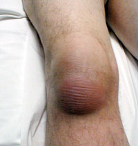
Clinical photo of infrapatellar bursitis, Courtsey: UCSD
Referring to diagnosis, Dr. Pinto says that the doctor will examine the knee for tenderness over the bursa. Sometimes, he/she may extract a fluid sample from the bursa, using a needle and syringe to check whether it is caused by an infection or is due to other causes. Blood tests and X-rays may also be carried out.
With regard to treatment, he says that the knee which is painful should be rested, propping it up on a pillow when sitting or lying down. It is important to remember not to put pressure on the sore and swollen knee until the swelling goes off and avoid irritation of the area.
An ice pack wrapped in a cloth and kept on the knee for about 20 minutes every 3 to 4 hours may also help to ease the pain. A knee sleeve or an elastic bandage could also be worn around the knee to reduce the swelling, he says, adding that the doctor may advise the patient to take anti-inflammatory medicine and apply an analgesic over the affected area.
Exercises as advised by the doctor will also help as also physiotherapy.


