Life-saving Ozaki procedure now in SL
By Kumudini Hettiarachchi

Dr. Muditha Lansakara
What is the link three women and one man from Hatton, Galagedera, Welimada and Pilimatalawa have with Ozaki?
All four of them, suffering from aortic valve disease, have undergone a pioneering procedure called the ‘Ozaki Procedure’ at the Kandy National Hospital over the past few weeks.
Introduced for the first time in Sri Lanka to deal with aortic valve disease in a different manner by a team led by skilled Consultant Cardiothoracic Surgeon Dr. Muditha Lansakara, the world had got this cutting-edge technique from Prof. Shigeyuki Ozaki of Japan about a decade ago.
Aortic valve disease causes shortness of breath and patients can suffer from complete dilatation (fully expansion or opening) of the heart, be prone to spells of giddiness as well as stroke. “If we leave them alone without an intervention, they can die,” says Dr. Lansakara.
Giving the medical name of the Ozaki procedure as ‘aortic valve neocuspidization with glutaraldehyde-treated autologous pericardium’, Dr. Lansakara says it can be performed for valve stenosis or regurgitation or a mix of both.
“The affected or diseased cusps are replaced by cusps formed from the patient’s own pericardium (the membrane enclosing the heart, consisting of an outer fibrous layer and an inner double layer of serous membrane). As such, it is ‘autologous’ and the chances of rejection are nil,” he points out.
 The surgical details follow – the patient is put to sleep (put under general anaesthesia), the chest is opened up through a midline sternotomy and 7X8 cms of the pericardium dissected.
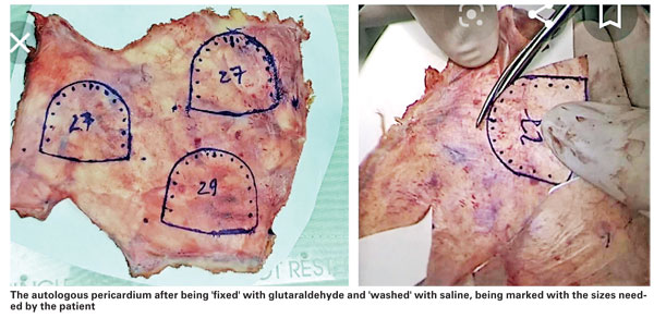
To allow the pericardium to be handled well, we ‘fix’ it by placing it in 0.6% of glutaraldehyde for 10 minutes after which we ‘wash’ it in saline thrice for six minutes each time, says Dr. Lansakara.
The patient, meanwhile, is cannulated and attached to the heart-lung (cardiopulmonary bypass) machine to allow for cardioplegia (the intentional and temporary cessation of the heart).
“Thereafter, we cut into the proximal aorta to reach the aortic valve. Depending on the pathology (disease) and requirement, the diseased cusps of the aortic valve are excised,” says Dr. Lansakara, explaining that all four patients had aortic valve stenosis, while three also had bicuspid valves, which means they had two instead of three cusps, caused by congenital calcification and narrowing of the aortic valve.
The skill and expertise come into play when the Cardiothoracic Surgeon has to mould or fashion three cusps from the harvested pericardium after getting the precise sizes needed by a particular patient, MediScene learns. These cusps are then fixed to the natural annulus of the aortic valve outlet which is ring shaped.
“The three commissures forming the three cusps of the aortic valve help the proper functioning of the valve in the most natural manner as it should have been, if not for the pathology. This prevents leaking,” says Dr. Lansakara.
Usually, what has been done in Sri Lanka before the Ozaki procedure is replacement of the diseased aortic valve with an artificial mechanical or tissue (biological) valve.
“Under this intervention, a ring is fixed to the annulus which, in fact, distorts the physiology of the aortic valve outlet, which is a 3-dimensional configuration. This distortion makes it mandatory for a blood-thinner (warfarin) to be prescribed and also to monitor patients with regular INR (international normalized ratio) tests,” says Dr. Lansakara, adding that if the INR is low there is the possibility of the patient’s blood clotting and obstructing the valve and if high there is a possibility of spontaneous bleeding. (The INR is a laboratory measurement of how long it takes blood to form a clot. It is used to determine the effects of oral anticoagulants on the clotting system.)
He says that as such patients who are engaged in manual labour or as carpenters etc may have to change their livelihoods and young women who are on warfarin have issues in having babies.
The bio-valve, meanwhile, used to have a short life but now there are newer long-lasting valves, but they are very expensive, it is learnt.
The advantages of the Ozaki procedure are that the patient’s own tissues are used, the 3-D configuration of the valve is maintained and the orifice of the aortic valve area is maximally used without narrowing. The patient gets only aspirin for about six months and not warfarin and there is no need for him/her to come into hospital for INR testing, it is learnt.
The cusps are moulded according to a set of ‘Ozaki sizes’ and the Cardiothoracic Surgeons have to be very skilled – with speed and precision being the need of the hour.
The trailblazing team which has introduced the Ozaki procedure to the country includes Consultant Cardiothoracic Surgeon Dr. Muditha Lansakara and Surgical Medical Officers Dr. Suranga Wijesinghe and Dr. Duminda Ratnasena; the Anaesthetic team headed by Dr. Priyantha Dissanayake who kept checking the pericardium valve competency through TOEs (transoesophageal echocardiograms – ultrasound scans of the heart using a special probe); Consultant Microbiologist Dr. Mahen Kotelawala who ensured that the glutaraldehyde was infection free; and the nursing team.
A special word of appreciation goes out from Dr. Lansakara to Priyanga from Sri Lanka Gamma who provided irradiation for the glutaraldehyde.
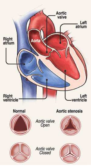
| All about the aorta The aorta is the largest artery in the body and begins at the top of the left ventricle, the heart’s muscular pumping chamber to take blood to all parts of the body. The heart pumps blood from the left ventricle into the aorta through the aortic valve. It is one of the two semi-lunar valves of the heart, with the other being the pulmonary valve. Altogether the heart has four valves and the other two are the mitral and the tricuspid valves. The aortic valve has three cusps or leaflets: the left coronary cusp (LCC), the right coronary cusp (RCC) and the non-coronary cusp (NCC). Aortic valve disease may be due to a condition present at birth (congenital heart disease) or other causes. Aortic valve disease includes: Aortic valve stenosis – here the cusps of the aortic valve get thickened and stiff or fused together. This will cause narrowing of the aortic valve opening and results in the valve not opening fully thus hindering the flow or blocking the flow of blood. This, in turn, will lead to blood flowing to the body being reduced or blocked. Aortic valve regurgitation – here the aortic valve does not close properly, causing blood to flow backward into the left ventricle. The signs and symptoms of aortic valve disease may include: Abnormal heart sound (heart murmur) when heard through a stethoscope Shortness of breath or fatigue particularly after a person has been very active or when he/she lies down Dizziness Fainting Chest pain or tightness Irregular heartbeat Not eating enough or not gaining enough weight, mainly in children Swelling of the ankles and feet | |


