News
Complex double heart surgery, a first in SL
View(s):- Team headed by Dr. Vivek Gupta performs ‘Extra anatomical bypass of coarctation
of the aorta’ along with ‘Mitral valve replacement’
By Kumudini Hettiarachchi
It started six months before with a small hathiya (panting) whenever he walked. Exhaustion also hit very fast.
Now not only Sisira Siriwardena but also his two mallis (younger brothers) are all smiles – this 41-year-old has been pulled back from the brink of death through a first in Sri Lanka.

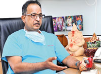
Dr. Vivek Gupta. Pix by M.A. Pushpa Kumara
Sisira is the first in the country to undergo a complex and complicated double open-heart surgery – the ‘Extra anatomical bypass of coarctation of the aorta’ along with ‘Mitral valve replacement’ at the Asiri Surgical Hospital in Narahenpita.
The team which ventured into this surgical field untried in Sri Lanka before was headed by Senior Consultant Cardiothoracic Surgeon Dr. Vivek Gupta.
Living and working in Habaraduwa and keeping the home fires burning for his wife and three-year-old daughter, the hathiya seemed very strange for active Sisira who was heavily into volleyball and cricket.
Doctors in Galle suggested he see a Cardiologist and that is what he did. He was then informed that there was a valve prashnayak (issue) in his heart.
However, after a second opinion and a plethora of tests including a repeat echocardiogram, angiogram and CT scan in Colombo that the distraught family realized that there were major problems in Sisira’s heart.
Immediate was the need to perform surgery and so he was admitted to hospital on March 12 with open heart surgery being performed on March 15.
Pacing the area outside the Operating Theatre (OT) as Sisira, their Loku Aiya, was wheeled in at 7.45 in the morning were Samantha and Gimhan.
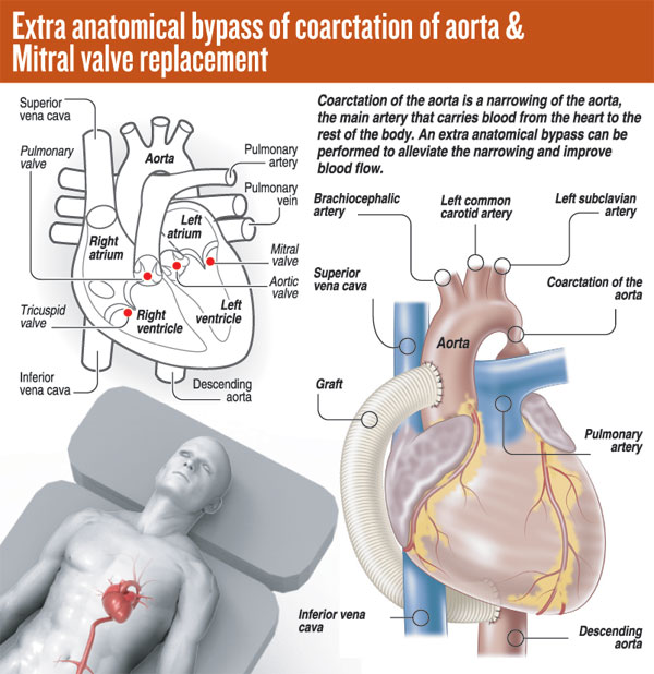
Dr. Gupta explains what ‘coarctation of the aorta’ means, before getting down to the technique and skill required to correct this major anomaly (See box).
“It is a birth defect where a part of the aorta is narrower than usual,” says Dr. Gupta, pointing out that in Sisira’s case, the narrowing was in a “bad place” –the aortic isthmus.

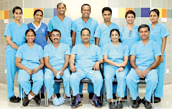
The team (seated) Senior Consultant Cardiothoracic Surgeon Dr. Vivek Gupta (centre) is flanked (from left) by Assistant Surgeons Dr. Chinthaki Suranimala & Dr. Kolitha Lelwala and Senior Consultant Anaesthetist Dr. Gayani Senanayake and Consultant Anaesthetist Dr. Asitha Dassanayake. Standing (from left) are Medical Officer of the Cardiothoracic Intensive Care Unit (CTICU) Dr. Andrea Harley; CTICU Sister-in-Charge H.M.C.J. Anuruddika; Theatre Technician Sanath Suraweera; Senior Perfusionist Pathum Chathuranga; Chief Perfusionist S.S. Senevirathna; Assistant Chief Nursing Officer & Sister-in-Charge of the Cardiac Operating Theatre (COT), GothamiWijayaweera; and COT Nursing Supervisor Wathsala Rajapaksha.
The aorta is the main artery that carries oxygen-rich blood from the heart to the rest of the body. The blood flows out of the heart through the aortic valve, passes through the aorta’s cane-shaped curve and supplies the other major arteries to deliver blood to the brain, muscles and other cells.
The aortic isthmus – like its literal cousin which is ‘a narrow strip of land with the sea on either side – is the segment of aorta located between the origin of the left subclavian artery and the connection of the ductus arteriosus to the descending aorta. (See graphic) “It is a difficult place,” concedes Dr. Gupta, adding that Sisira also had another issue – a heavily calcified mitral valve. This is the valve between the two chambers (atrium and ventricle) of the heart’s left side.

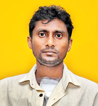
Sisira Siriwardena
The Sunday Times understands that in the first instance Sisira’s condition had been diagnosed as just rheumatic mitral valve disease and he could easily have died on the operating table if he had undergone surgery for that repair alone.
Later, after correct diagnosis, when the situation was explained to the desperate family and they turned to Dr. Gupta with pleading eyes, he had told them of the risks involved but that “it can be corrected”.
When the Sunday Times meets the three brothers, Sisira has been discharged on March 25 and they are at the hospital for a follow-up visit. The smiles of relief are very wide.
“Den sahalluwak thiyenawa (Now there is no stress),” says Sisira, explaining that he is on a healthy lifestyle regimen of diet and exercise along with just one tablet.

| Intricate steps in this single-stage correctionThe surgical steps for ‘One-Stage Relief of Aortic Coarctation with Mitral Valve Replacement’ as detailed by Dr. Vivek Gupta were:n Having placed Sisira under general anaesthesia, the team had first exposed the groin’s femoral artery and vein as these two vessels which start in the upper thigh are a continuation of the external iliac artery and vein. These were used to supply blood to the lower part of the body including the kidneys because from normal aortic cannulation (needed for cardiopulmonary bypass), the lower body would not get blood due to the aortic coarctation. This was why the team cannulated both the aorta and the femoral artery n Next, Sisira’s chest (sternum) had been opened up and he had been put on the heart-lung machine. n Emptying Sisira’s heart of its blood, the ‘starfish’ apparatus had been used to lift the heart out of the chest cavity (retract). n The team had then cut open the layer behind the heart to find the thoracic aorta. Using a side clamp on the thoracic aorta, they had made a small opening, suturing (stitching) a size 14 Dacron (woven) allograft onto it. The graft had been taken from around the back of the heart to the front. n Next was the mitral valve replacement. Cross-clamping the top of the aorta (ascending aorta), cardioplegia (a solution to temporarily stop the heart from beating) had been administered. Thereafter, the team had opened up the heart’s right atrium and through that incision (cut), accessed the atrial septum (the wall separating the right and left atria of the heart). This too they had opened up to get to the mitral valve. This valve had been heavily calcified with only a small hole remaining for blood flow. n The next step had been to excise (remove) carefully, using a pair of scissors, the diseased mitral valve. As the area for the insertion of the new valve was very small, only a 21-size valve would have been suitable. However, as no 21-size mitral valves were available in the market, the team had innovated by using a 21-size aortic valve in reverse position (as the blood flows the other way). This valve had been put in place with ethibond sutures. The septum and right atrium had then been closed, after checking the valve. n The final steps had included connecting the top of the graft to the aorta, bypassing the narrowed section – placing a side clamp on the top of the aorta and suturing the top of the allograft to it. n Taking Sisira off the heart-lung machine, two drains had been placed along with pacing wires. The chest wall had thereafter been closed using stainless steel wires, as usual. It was a tense three-hour open heart surgery but it took about 5½ hours because the tissues were diseased and there was a lot of bleeding vessels here and there, adds Dr. Gupta. | |
The best way to say that you found the home of your dreams is by finding it on Hitad.lk. We have listings for apartments for sale or rent in Sri Lanka, no matter what locale you're looking for! Whether you live in Colombo, Galle, Kandy, Matara, Jaffna and more - we've got them all!

